| Fundus Perimetry | |
|---|---|
| Projection field | 30° (radius) |
| Background luminance | 31.4 asb |
| Maximum luminance | 10000 asb |
| Dynamic range | 0 – 50 dB |
| Stimulus size | Goldmann III (26”) |
| Stimulus duration | 200 ms |
| Threshold tests | 24-2, 10-2 |
| Fixation control | 25 Hz automated retinal Tracking |
| Foveal threshold testing | |
| Automatic pupil measurement |
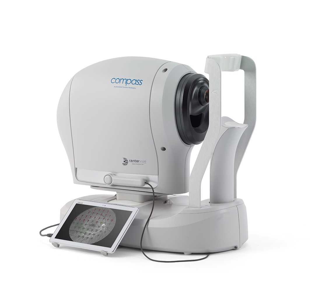
iCare COMPASS Automated Perimeter with active Retinal
Original price was: $13,999.00.$7,999.00Current price is: $7,999.00.-43% OFF
iCare COMPASS provides confocal retina images combined with an automated perimeter, active retinal tracking function, and a scanning ophthalmoscope.
In stock
Description
iCare COMPASS device is a non-mydriatic scanning ophthalmoscope that provides confocal images of the retina combined with an automated perimeter. Different visual fields such as 24-2, 10-2, and 30-2 can be obtained with other strategies, as well as True Color, Red-Free, and IR images.
iCare COMPASS overcomes the limitations of Standard Automated Perimetry (SAP). The automatic perimeter has an active retinal tracking function and a scanning ophthalmoscope, which measures retinal sensitivity without artifacts caused by eye movements, confocal TrueColor fundus images, and fixation analysis.
- iCare Compass combines the field of view measurement, active retinal tracking that corrects loss of fixation, and confocal TrueColor fundus photography.
- Ease of use thanks to non-mydriatic, auto-focus (no need for test tubes), touch screen, automatic adjustment, and easy cleaning routine.
- Patient-friendly: simple and fast tests that can be stopped and restarted along the way without losing measurement data.
- Fully compatible with SAP; standard 24-2, 30-2, and 10-2 field of view measurement with age-adjusted database, super-threshold value, and quick super-threshold value measurement.
- Active compensation for eye movements by continuous, automatic tracking of eye movements ensures an accurate correlation between function (threshold value) and structure (fundus image).
- iCare COMPASS, with its built-in fundus camera, takes 60 ° confocal images: TrueColor, infrared and red-free. In addition to 3D visualization of optic nerve head (ONH).
- Browser-based software: remote, password-protected, access to measurements and images for all PCs in the same local area network (LAN).
- Print reports: survey and progression reports
What advantages does iCare COMPASS have?
One of the significant advantages of the iCare COMPASS automated perimeter is that it can measure the retina’s sensitivity in specific places; it has more precision thanks to the compensation based on retinal tracking of eye movement and a simultaneous evaluation of the function and the structure of it.
This device overcomes the limitations of standard automated perimeters for the clinical management of glaucoma.
The iCare COMPASS is a scanning ophthalmoscope combined with an automatic perimeter.
‘Fundus automated perimetry’ is a technique that describes the retina during a visual field test, and this allows a correlation between vision and retinal structure. The advantages of this technique over a standard automatic field of view test are the ability to measure sensitivity in a specific part of the retina, the greater accuracy achieved by compensating eye movements based on retinal monitoring, and the simultaneous evaluation of eye function based on retinal sensitivity and structure.
iCare COMPASS expands the field coverage for the first time by 30 ° + 30 ° and uses luminance parameters and a sensitivity scale as in a standard automatic field of view test.
Product features
The iCare COMPASS fundus tracking automatic perimeter is the only auto-perimeter that goes beyond the functional limits of traditional auto-perimeters.
iCare COMPASS combines an automated perimeter with active retinal tracking and a fundus scanner to
detect artifact-free retinal sensitivity due to eye movements, analyze fixative conditions, and
capture TrueColor confocal retinal images.
1. Visual Field Test

Compatibility with standard automatic perimeter
Automatic visual field tests with iCare COMPASS provide full compatibility with standard 24-2, 30-2, and 10-2, and these visual field tests include sensitivity measurements, including an age-friendly normal eye database. To do.
The iCare COMPASS splash hold test is used for fast screening to detect visual field defects.
2. Active Retina Tracking
![]()
Improved reliability when fixation is poor
Retina tracking is the most important feature of fundus tracking automatic perimeter.
Retina tracking is always automatic, tracking active eye movements and correcting fixative shifts in real-time. Immediately move the target position of the retina.
This active retinal tracking can accurately correlate function (retinal threshold) with structure (fundus image).
You can also increase the reliability of changes in the visual field over time.
3. Can be used with simple operation

No trial lens required
Since the conventional dome-shaped perimeter is performed by refraction correction using a trial lens, an optional lens is required. Also, lens artifacts can occur. iCare COMPASS is equipped with an autofocus function without using any trial lens, which shortens the inspection time and improves usability.
The test results may change depending on the trial lens for dome-shaped perimeters.
- If the lens is too close, rubbing the eyelashes will make you worried about the presence of the lens, and you will not be able to concentrate.
- If the lens is too far away, the frame of the lens may block the field of view, causing ring-shaped dark spots.
- If the lens position is misaligned, it may cause partial loss.
- If you use a face mask, the trial lens may become cloudy.
4. TrueColor Confocal Fundus Imaging
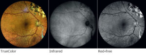
Improved diagnosis and prognosis in glaucoma management
Evaluation of the fundus helps in the precise diagnosis of glaucoma.
iCare COMPASS is the first visual field test to provide 60 ° TrueColor, infrared and Red-free confocal fundus images. Displays images of the retina in various modalities.
iCare COMPASS Technical Specifications
| Fundus Imaging | |
|---|---|
| Field of view | 60° (diameter) |
| Bi-focal Stereo Image of the ONH | |
| Sensor resolution | 5 Mpixel (2592×1944) |
| Light source | infrared (825-870 nm) and white LED (440-650 nm) |
| Imaging modalities | color, infrared, red-free |
| Resolution | 17 microns |
| Dimensions | |
|---|---|
| Weight | 25 Kg |
| Size | H 620 X W 590 X D 360 mm |
| Electrical requirements | |
|---|---|
| Power | 100-240 VAC, 50-60 Hz |
| Consumption | 80 W |
| Other Features | |
|---|---|
| Automatic operation | auto-alignment, autofocus, auto-retinal tracking, auto-pupil tracking, auto-exposure, auto-capture |
| Non-mydriatic operation | minimum pupil size 3 mm |
| Working distance | 28 mm |
| Auto-focus range | -12D to +15D |
| Fixation target | programmable, internal |
| User interface | Tablet operated, with multi-touch, color display |
| Connectivity | Ethernet connection through the device |
| DICOM support | modality worklist |
| Hard disk | modality worklist |
Reference Library
Only logged in customers who have purchased this product may leave a review.
Reviews
There are no reviews yet.