


Haag-Streit Octopus 600 Pro Computer Perimeter
Original price was: $13,888.00.$7,888.00Current price is: $7,888.00.-43% OFF
Octopus 600 is used to detect the initial stage of glaucoma development by combining the Pulsar method for early glaucoma detection
In stock
Description
Octopus 600 is a computer perimeter that analyzes contrast sensitivity. It is used to detect the initial stage of glaucoma development. Pathological disorders can be identified even when SOCT and IOP parameters are within the normal range. Thanks to innovative technology, diagnostics are only 2-4 minutes long.
The Octopus 600 extends the successful HAAGSTREIT product range of visual field analyzers combining the Pulsar method for early glaucoma detection and standard white-on-white perimetry for long-term follow-up in one compact and standalone device. It is the first visual field analyzer that performs standard white-on-white perimetry on an integrated flicker-free screen, using TFT and LED technology.
The Haag Streit Octopus 600 perimeter features new Pulsar early detection and standard white-on-white perimetry, with LED technology and a touch screen. This instrument is compact-sized with simplified perimetry to streamline operation and provide a range of visual field analyses.
Octopus 600 Features:
-
Central Field Static Perimetry
In the central visual field, the Octopus 600 performs standard white-on-white threshold testing in just 2–4 minutes. Its comprehensive test library for main tests, including G, 32, 30-2, 24-2, M, and 10-2, and its flexible printouts in Octopus and HFA format cover your essential clinical needs.
-
Testing
Offers the most commonly used static tests. For central field testing, there are the physiology-based G-patterns following the retinal nerve fibers and the 32, 30-2, and 24-2 patterns. For the macula, there are the physiology-based M pattern and the 10-2 pattern. With the fast TOP strategy, full threshold testing can be completed in 2-4 minutes.
-
Fast Screening
Use your Octopus 600 as a fast screening device and quickly distinguish between normal and abnormal visual fields. Screening can be performed with standard white-on-white or the patient-friendly Pulsar perimetry designed for early glaucoma detection.
-
Glaucoma Screening
Use the new Glaucoma Screening Test GST to distinguish between normal and abnormal visual fields in less than one minute. The test is purely qualitative and distinguishes between normal and abnormal visual fields by presenting stimuli three times at a brightness that patients with normal vision should see. If not seen, the visual field is flagged as abnormal with high reliability, and the patient can be further tested. Because the test is efficient, it opens doors for more routine visual field testing to ensure no pathology goes undetected.
-
EyeSuite
Immediately identify levels of change with the EyeSuite Progression Analysis. It not only reveals whether change is significant but also whether it is local or diffuse and how fast the change was happening. For an effective clinical workflow, all results are displayed using intuitive graphical symbols and can be viewed directly in your office if the Octopus is networked.
-
Easy Integration
The EyeSuite software has been designed for optimum patient flow in busy practices. It controls all Haag-Streit devices and allows them to be networked with other Haag-Streit devices, your office computer, and your EMR system without needing any expensive third-party software.
-
Standard 7-in-1 Printout
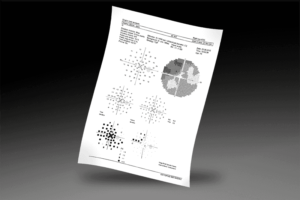
Look at visual field results the way you are used to from your Octopus perimeter. All Octopus perimeters offer the standard 7-in-1 Printout with its well-known representations, a customizable 4-in-1 printout, a serial printout, and much more. And why not conveniently view results in your office by networking your Octopus to the EyeSuite software on your computer?
-
Cluster Analysis
The sensitive Cluster Analysis groups visual field defects along nerve fibre bundles and combines high sensitivity with reasonable specificity to detect early glaucomatous changes. Significant defects are highlighted, and the cluster defects can be used for structural comparison. Cluster Analysis is available in both single field and trend view.
-
Polar Analysis/Trend
Combining the results of both structure and function is key to obtaining a comprehensive assessment of the onset and progression of glaucoma. The Octopus Polar Analysis projects local visual field defects along the nerve fibers to the optic disk and displays them oriented as structural results. This makes structure-function correlation almost intuitive. Polar Analysis is available in both single field and trend view.
-
Fixation Control
Automatically eliminate fixation losses from your visual field testing.
STANDARD TEST LIBRARY
The central tests you need
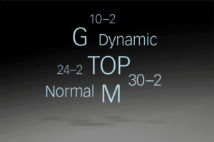
The Octopus 600 offers the most commonly used static tests. For central field testing, there are the physiology-based G-patterns following the retinal nerve fibers and the 32, 30-2, and 24-2 patterns. For the macula, there are the physiology-based M pattern and the 10-2 pattern. With the fast TOP strategy, full threshold testing can be completed in 2-4 minutes.
OCTOPUS PRINTOUT
Standard Octopus representations

Look at visual field results the way you are used to from your Octopus perimeter. All Octopus perimeters offer the standard 7-in-1 Printout with its well-known representations, a customizable 4-in-1 printout, a serial printout, and much more. And why not conveniently view results in your office by networking your Octopus to the EyeSuite software on your computer?
HFA-STYLE PRINTOUT
Smooth transition from HFA
Enjoy a smooth transition from an HFA to an Octopus perimeter. All Octopus perimeters allow you to import your historic HFA data. Because raw data is imported, you can display your historical and new data in the same format of your choice, either as an HFA-style printout or in the Octopus format. To learn more about how the two formats correlate, watch the movie.
EYESUITE GLAUCOMA ANALYSIS
A clear view of glaucoma

Get the most out of your glaucoma visual field with the highly sensitive Cluster Analysis, the intuitive Polar Analysis for structural comparisons, and the easy-to-interpret EyeSuite Progression Analysis.
CLUSTER ANALYSIS/TREND
Sensitive glaucoma analysis

The sensitive Cluster Analysis groups visual field defects along nerve fibre bundles and combines high sensitivity with reasonable specificity to detect early glaucomatous changes. Significant defects are highlighted, and the cluster defects can be used for structural comparison. Cluster Analysis is available in both single field and trend view.
Main advantages of Haag-Streit Octopus 600 Pro automatic computer perimeter
The Haag-Streit Octopus 600 Pro Automatic Computer Perimeter has better features than the base model. Thanks to cluster analysis, you can process survey data in an extended software range. Key changes include the TOP Optimized Faster Threshold Strategy, which increases inspection speed by up to 80%, with standard W/W perimetry. The study is now an order of magnitude faster while maintaining the highest accuracy of the topography of spatial contrast sensitivity.
Otherwise, the Haag-Streit Octopus 600 Pro specialized automatic computer perimeter retained all the advantages of the primary device. The weight and dimensions of the rig have remained the same despite improved features and software.
The built-in video camera is responsible for the continuity of the examination, and the generation of stimuli takes place through a TFT screen. The binocular system has a set of corrective lenses ranging from -8 to +4 D.
Specifications:
- Stimulus generation type: TFT monitor
- Field of View: 0⁰ to 30⁰
- Fixation control: integrated video camera
- Types of perimetry: standard W/W; pulse (Pulsar)
- Characteristics of the Pulsar technique: background 32 cd/m2; stimulus range ~35 src; stimulus size 5; stimulus duration 500 ms at a frequency of 10 Hz; Gp and 32p programs; compatible with TOP strategy
- Characteristics of standard perimetry W/W: background 10 cd/m2; stimulus range ~35 dB; stimulus size 0.43 (Goldman III); stimulus duration 100 ms; programs G, 32 and 24-2; compatibility with threshold and dynamic strategies, as well as with the TOP strategy
- Software: threshold schemes of the center and periphery; cluster and polar analysis; progression analysis; anatomical program G-program
- Perimetry Strategies: TOP (Optimized Accelerated Threshold Strategy to speed up examination up to 80%); threshold; dynamic
- Ability to output data by the Humphrey Protocol (HFA)
- Interfaces: 4 USB 2.0; 1 Base-T 1000
- Power: 100-240VA; 50/60 Hz
- Instrument dimensions: 467*508*500 mm
- Weight: 12.7 kg
Include:
- TABLE
- Power cable and clicker.
- Unit include manufacture lenses (+1.0,+2.0,+3.0,+4.0,-1.0,-2.0,-3.0,-4.0,-5.0,-6.0,-7.0,-8.0)
Reference Library
Only logged in customers who have purchased this product may leave a review.
Reviews
There are no reviews yet.