
NIDEK Mirante SLO/OCT Multimodal Imaging Platform
Original price was: $30,999.00.$20,999.00Current price is: $20,999.00.-32% OFF
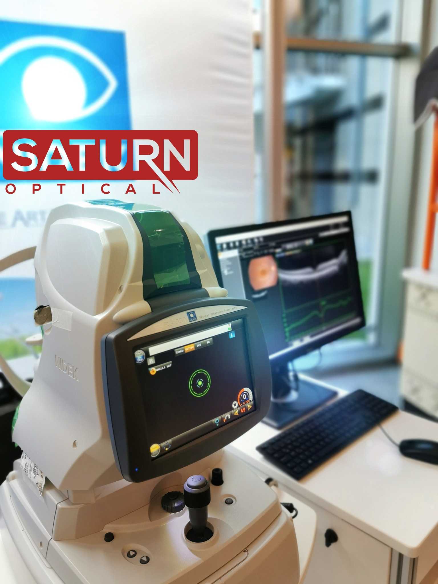
NIDEK Mirante is equipped with a high-resolution 4K technology system, a scanning laser ophthalmoscope, and the ideal image of the anterior segment of the eye and the fundus.
In stock
Description
NIDEK Mirante is An ideal image of the anterior segment of the eye, and the fundus is the most important part of an ophthalmological examination. The most powerful device for this purpose is the Nidek Mirante optical coherence tomography.
NIDEK Mirante is equipped with a high-resolution 4K technology system and a scanning laser ophthalmoscope, which allows you to use the ultra-modern pSLO technique. The device functions as a retinal fundus camera equipped with advanced laser angiography mode.
The SLO Mirante system makes it possible to detect a significantly greater number of elementary lesions than the RNM in the 7 ETDRS fields and visualize peripheral lesions using ultra-widefield imaging. THEREFORE, the SLO Mirante system is efficient and suitable for clinical practice screening and monitoring diabetic retinopathy.
NIDEK Mirante Features
- The ultimate multimodal imaging platform
For the SLO/OCT model
– Color / FA / ICG / Blue-FAF / Green-FAF / Retro mode
– OCT / OCT-Angiography*
For the SLO model
– Color / FA* / ICG* / Blue-FAF /Green-FAF / Retro mode - Ultra wide-field x ultra HD image*
- Unsurpassed color
- Dynamic/Simultaneous FA and ICG
- Unique Retro mode
- HD wide-area OCT
- Fly Through function
Specifications:
- OCT Technology: Optical Coherence Tomography
- pSLO technology: image reconstruction using a scanning laser ophthalmoscope; 3 simultaneous laser wavelengths for color fundus imaging in IR mode
- Laser angiography mode: 4 laser wavelengths for FA and ICG modes; algorithms for quantitative analysis of changes in blood vessels; panoramic scan function
- 880nm IR Laser Enhanced Wide Angle Imaging Function
- Powerful 4K HD Digital Imaging System
- A high degree of automation: automatic system for positioning and focusing the eye; automatic tracking in the center of the pupil
- Axial resolution: 3 µm
- Scanning speed: 85000 A-scans per second
- Minimum pupil diameter: 2.5mm
- Possibility of using the system as a retinal fundus camera
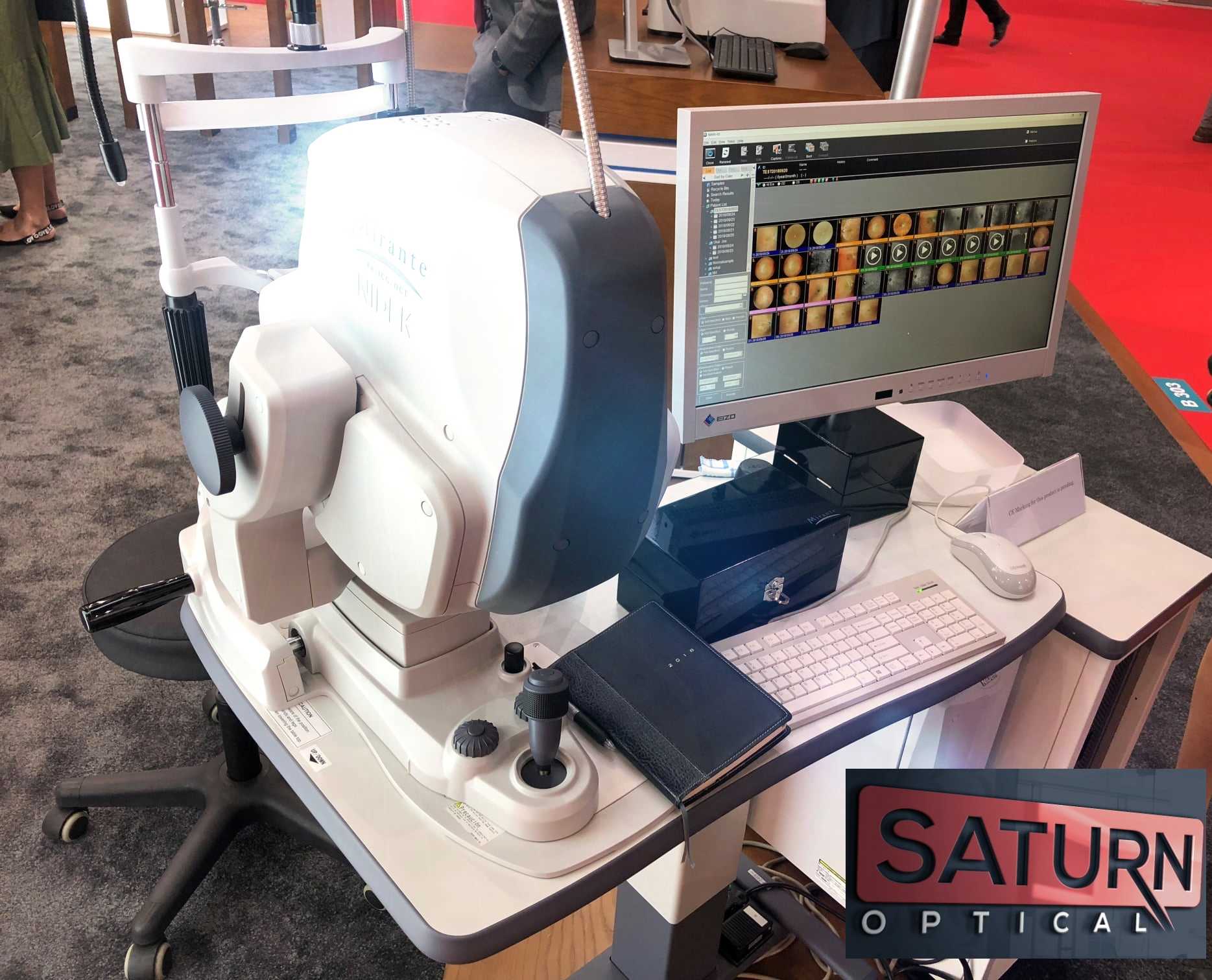
- Ability to freely transfer visual data to a computer or laptop specialist
- A software package, including NAVIS-EX for comprehensive setup and management of various studies, as well as multiple protocols for diagnostics using multifunctional data display
Nidek Mirante Advantages
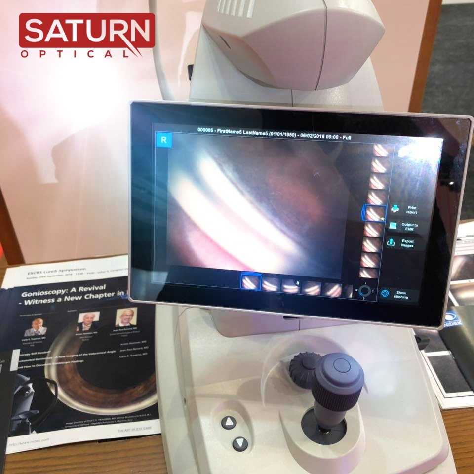
The stunning image quality obtained using the 4K HD digital system clearly distinguishes the Nidek Mirante, optical coherence tomograph from other devices in its class. This unique device uses a scanning laser ophthalmoscope to obtain high-precision images of the fundus, retina, and anterior segment of the eye, followed by data processing on a doctor’s personal computer or laptop. In addition, the unit works as a fundus camera, which will save you both budgetary funds and space in your office.
Optical, digital, and software tools give the Nidek Mirante confocal coherence tomography a major advantage in more complex examinations, such as during laser angiography procedures. The pupil size minimum suitable for starting any analysis using the device is two and a half millimeters. In most clinical cases, you do not need to use medication on the patient’s eye to begin the examination.
High Definition of cSLO and OCT wide imaging system. All in one device that includes all functions necessary for retinal diagnosis.
Colour SLO
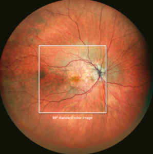
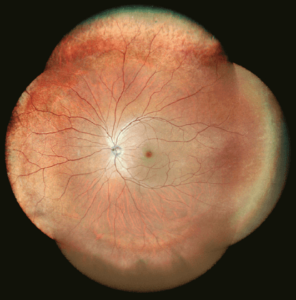
163º ultra wide-field image enables detailed evaluation of pathologies from the fovea to the extreme periphery.
Panorama image composition
Preset fixation points capture details of pathology even in the extreme periphery.
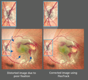
Flex Track Technology
The NEW flex track algorithm corrects image distortion due to the instable fixation and enhances averaging quality.
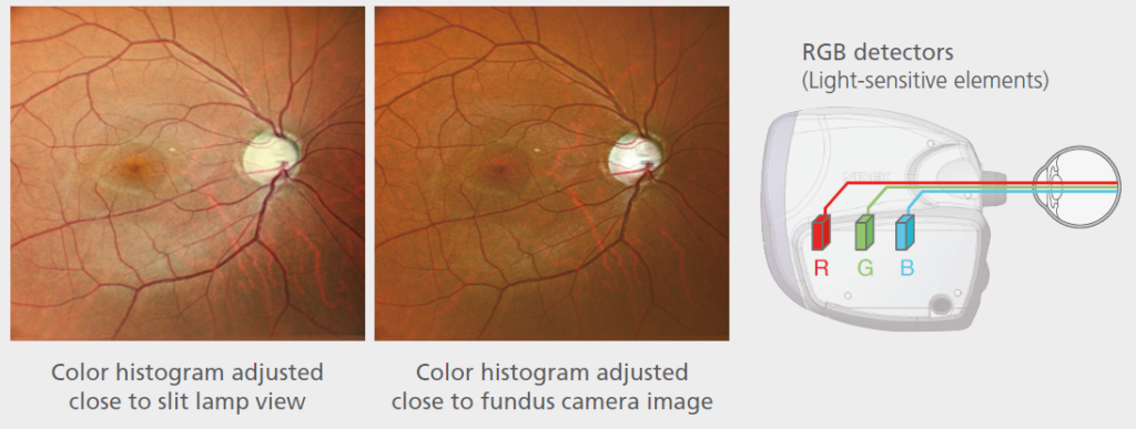
RGB triple detectors
Three separate RGB detectors simultaneously scan different depths of the retina with red, green, and blue wavelengths.
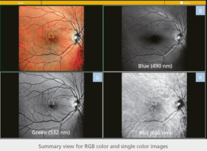
RGB color & selectable color display with a single slot
Single-color images in red, green, and blue wavelengths can be displayed after color image acquisition.
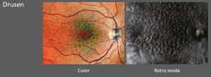
Retro Mode
A unique non-invasive technique for detecting pathologic changes in the choroid
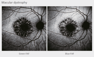
Blue FAF/ Green FAF fundus autofluorescence
FAF imaging is a non-invasive method to evaluate the retinal pigment epithelium (RPE) without contrast dye.
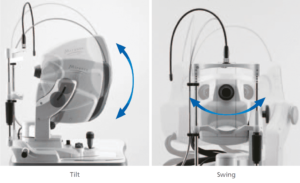
Easy to use functions
Simple interface and straightforward operation with tilt and swing features
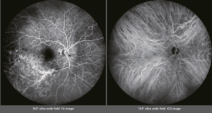
163º ultra wide-field FA & ICG images
Ultra wide-field imagining is available with the optional wide-field adapter.
HD dynamics and static angiogram
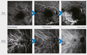
Auto gain control (AGC) optimizes gain level and contrast for early, peak, and late phases of angiography.
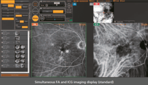
Simultaneous FA and ICG
The Mirante allows simple, simultaneous acquisition of FA and ICG images.
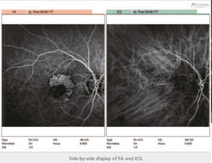
Easy Comparison of FA and ICG
The viewer software can present FA and ICG images side-by-side. An easy comparison is helpful for comprehensive evaluation.
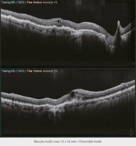
HD wide-area OCT
The maximum 16.5 x 12 mm area scan available with the Mirante allows wide-area diagnosis, including the macula and optic disc in a single shot.
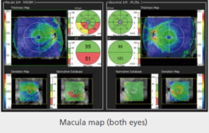
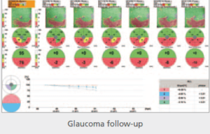
Glaucoma Analysis
The Mirante incorporates a 16.5 x 12 mm thickness map which visually presents pathological changes from the central retina to the periphery.
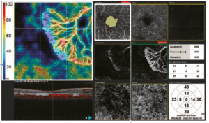
Anglo Scan – Segmentation into multiple tabs
The simple interface provides seven slabs for the macula map/ four slabs for the disc map with intuitive functionality and removal of projection artifacts:
– Vessel density map and perfusion density map
– Auto-detection of FAZ and shape analysis
– Wide area scan
– Tracing HD plus
– Selectable Definition
Reference Library
Only logged in customers who have purchased this product may leave a review.
Reviews
There are no reviews yet.