

TOMEY TFC-1000 Non-Mydriatic Fundus Camera
Original price was: $7,520.00.$4,520.00Current price is: $4,520.00.-40% OFF
Explore the TOMEY TFC-1000 Fundus Camera, a state-of-the-art ophthalmic device for efficient and accurate eye examinations. Discover its features like automatic eye tracking, non-mydriatic imaging, and a 12M pixel sensor for detailed fundus analysis. It is ideal for diagnosing and monitoring retinal diseases.
In stock
Description
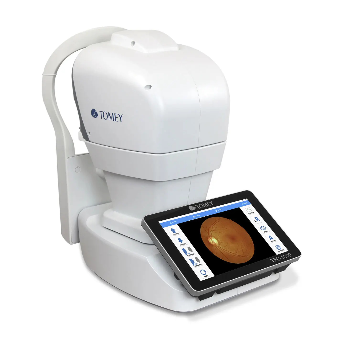
The TOMEY TFC-1000 is a fundus camera with several advanced features. It is designed to capture images of the fundus, the eye’s interior surface opposite the lens, which includes the retina, optic disc, macula, fovea, and posterior pole.
The TFC-1000 fully automatically generates a fundus image within 15 seconds using eye tracking, auto-focus, and auto-measurement. The “KISS” (Keep It Short & Simple) principle had the highest priority in the software and workflow design. This makes the operation of the TFC-1000 extremely easy to handle. The user-friendly interface is very intuitive and provides the results in a very short time.
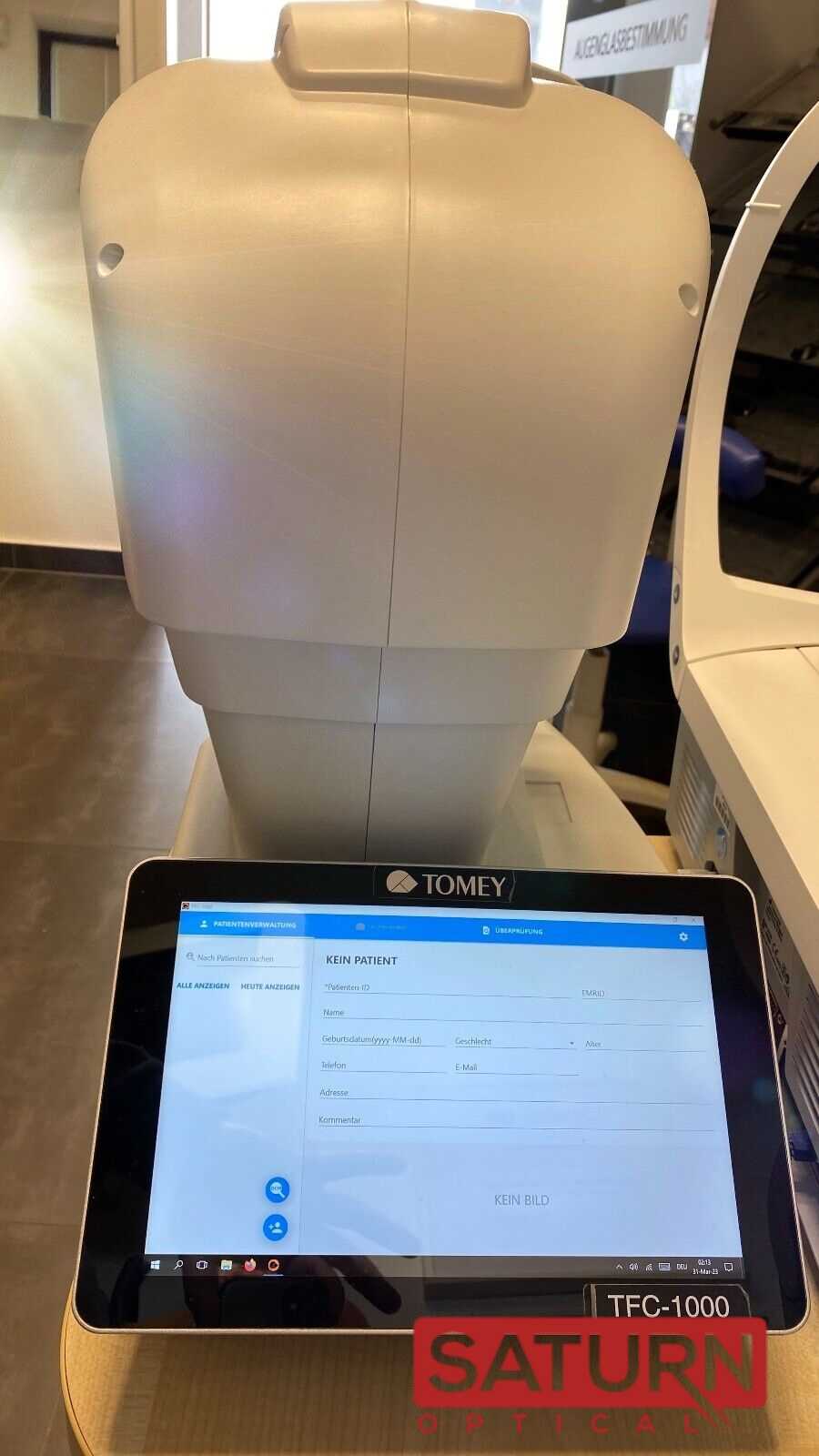
This principle was also used to get a streamlined hardware with built-in 10.1” true colour touch screen and all-in-one PC. Win10 as Operation System and the modern connectivity will fit easily in your clinical environment.
The TFC-1000 features:
- Automatic Eye Tracking and Auto Measurement: The camera can automatically track the eye and focus, enabling quick and accurate measurements.
- Rapid Image Acquisition: It can generate a fundus image within approximately 15 seconds, making the process fast and efficient.
- High-Quality Imaging: The TFC-1000 is equipped with a 12-megapixel sensor, ensuring high-resolution images for detailed examination.
- Wide Field of View: The camera offers a field of angle of 45° x 45°, which allows for a comprehensive view of the fundus.
- Multiple Fixation Points: It has nine fixation points, which aid in capturing different areas of the retina for a thorough examination.
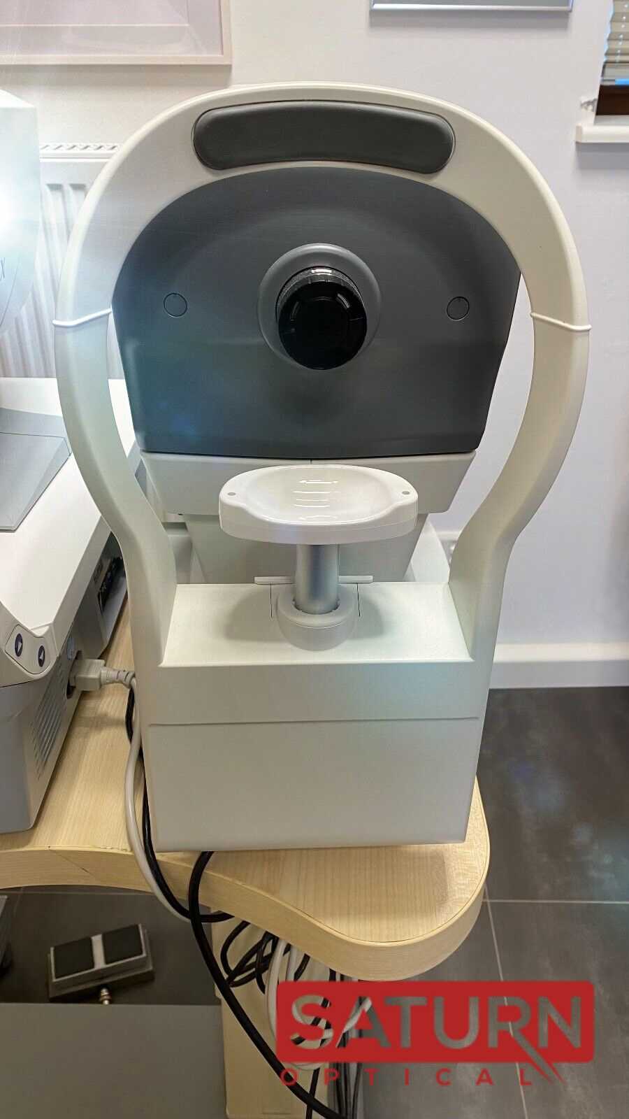
- Non-Mydriatic Camera: This feature indicates that the camera can take fundus images without the need for dilating the pupils, which is more comfortable for patients.
- Built-In Stitching Module Mosaic: This feature likely aids in creating a composite image of the fundus by stitching together multiple images.
- Anterior Segment Imaging: Besides the fundus, the camera is also capable of imaging the anterior segment of the eye.
- User-Friendly Design: The TFC-1000 was designed with simplicity in mind, following the “KISS principle” (keep it short and simple), making its operation straightforward and easy to handle.
- Connectivity: The camera includes WiFi functionality, enhancing its usability and ease of data transfer.
This camera appears to be a sophisticated tool for ophthalmologists and optometrists, providing them with a quick and detailed view of the eye’s interior for diagnostic purposes.
The TOMEY TFC-1000 fundus camera is used primarily in the field of ophthalmology and optometry for the following purposes:
- Diagnosing Eye Diseases: It helps diagnose various eye conditions affecting the retina, optic disc, and other parts of the fundus. Diseases like diabetic retinopathy, glaucoma, macular degeneration, and retinal detachment can be identified through fundus imaging.
- Monitoring Disease Progression: For patients with known retinal diseases, the TFC-1000 can be used to monitor the progression of the disease over time. This is crucial in managing chronic conditions and assessing the effectiveness of treatments.
- Screening for Eye Conditions: In routine eye examinations, the TFC-1000 can be used to screen for early signs of eye diseases, even before symptoms become apparent. This early detection is important for preventing or slowing down the progression of eye diseases.
- Documenting Changes in the Eye: The high-resolution images captured by the camera serve as a record of the current state of the eye, which can be valuable for comparing changes over time.
- Enhancing Patient Consultations: The images produced by the camera can be used to explain conditions to patients, helping them understand their eye health better.
- Research and Education: In academic and research settings, the TFC-1000 can be used to study the fundus in detail and contribute to medical research and education in the field of ophthalmology.
The non-mydriatic feature of the TFC-1000, which allows for imaging without pupil dilation, makes it more comfortable for patients and more convenient in clinical settings. The camera’s automatic features, such as eye tracking and auto-focus, streamline the imaging process, making it efficient for both the practitioner and the patient.
Only logged in customers who have purchased this product may leave a review.
Reviews
There are no reviews yet.