Huvitz HOCT-1F Retinal OCT with Fundus Imaging
Original price was: $25,999.00.$15,999.00Current price is: $15,999.00.-38% OFF
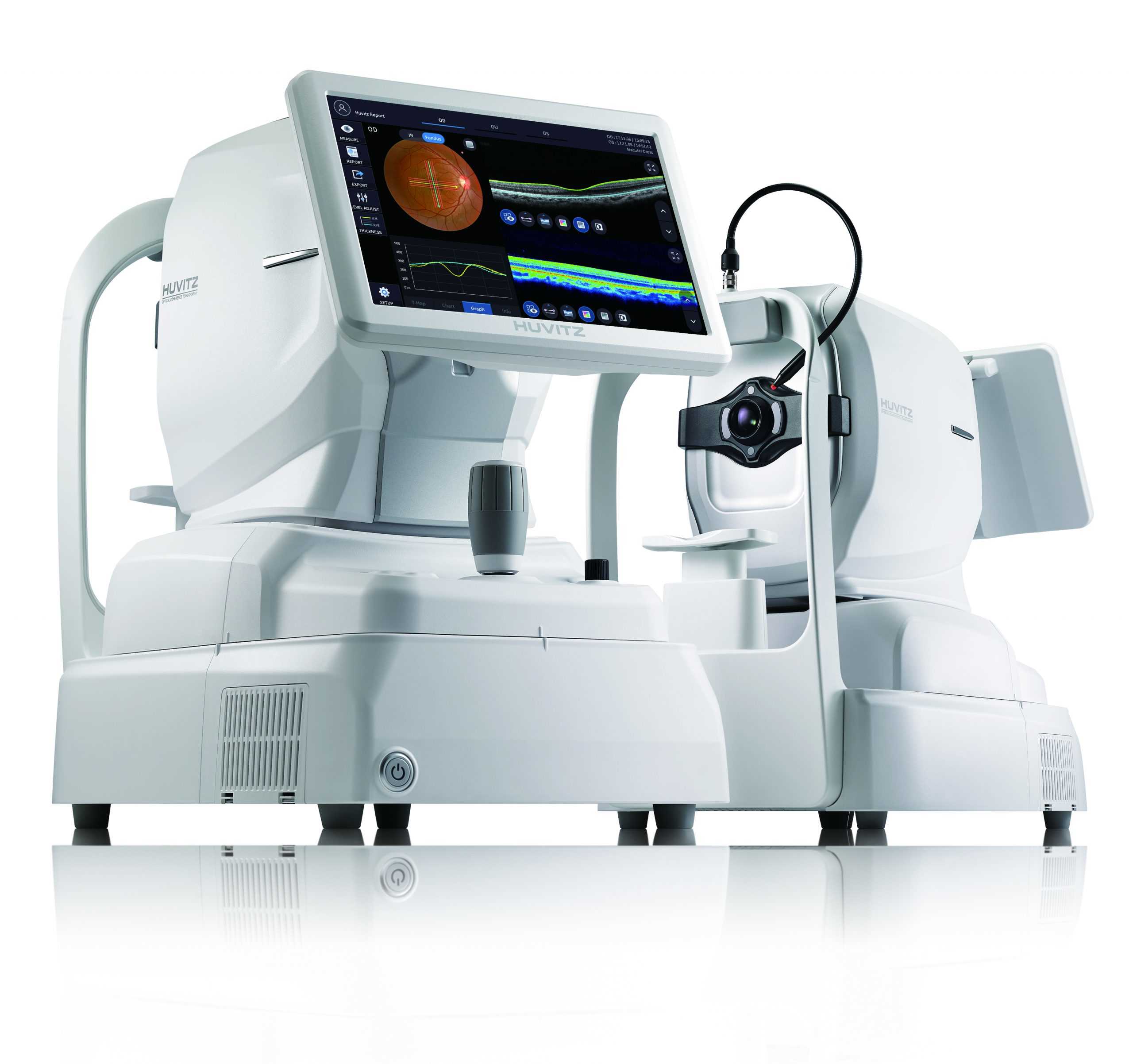
Huvitz HOCT-1F Offers high-speed scanning and high-quality imaging through the use of Huvitz’s outstanding optical technology and innovative imaging software. It combines modern OCT functionality and a compact, ergonomic body.
In stock
Description
Huvitz HOCT-1F is a one-of-a-kind in One HIGH SPEED AND HIGH QUALITY. Offers high-speed scanning and high-quality imaging through Huvitz’s outstanding optical technology and innovative imaging software.
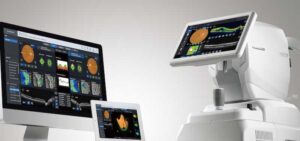
The all-in-one HOCT is easy to use. With one button, it creates a high-speed scan and a high-quality image, offering an excellent perspective for the ophthalmology clinic. One slot allows for capturing various types of analysis and diagnostic results. Easy to use, and the results are prominent and easy to follow. The all-in-one HOCT Huvitz will be the icon to lead a new era of Optical Coherence Tomography (OCT).
The new multimodal optical coherence tomograph from Huvitz is the result of many years of development and analysis of the needs of the ophthalmic market. It managed to combine modern OCT functionality and a compact, ergonomic body.
Huvitz All-in-One Optical Coherence Tomography (OCT)
Combining several functions, the device allows you to obtain various types of analysis and diagnostic results. Easy to use and has excellent results that are easy to interpret. The Huvitz OCT All-in-One will become an icon to usher in a new era of Optical Coherence Tomography (OCT).
The HOCT-1F integrated fundus camera offers an intelligent all-in-one solution for your screening offer. The auto-tracking, as well as the automatic triggering, make the operation very easy.
The 68,000 A-scans per second deliver high-resolution images in a concise time. In this way, abnormalities in the retinal structures can be detected. Signs of eye diseases such as AMD, glaucoma, or diabetic retinopathy are often difficult to detect on fundus-only images.
With the HOCT, you can check the individual layers for abnormalities. The additional software HOCT-Viewer for an external PC is optionally available, which enables you, for example, to display 3D. An anterior module for the HOCT is available separately.
Huvitz HOCT-1F Features:
- High speed
- Accurate and stable image averaging
- Retinal layers vividly visualized
- Adjust intensity level
- Combined in One
- compact design
- web search system
- Fast and stable automatic mode
- semi-automatic mode
- Large scanning area
- Innovative scan technology with motion detection technology
- Provides varied and functional scan patterns
- Progression to track pathological changes
- Compare patient symptoms before and after
- High-speed, large-area 3D modeling
- OU for binocular cross-analysis function
- Overview: Monocular Scanning and OCT / Fundus Imaging
- Detailed Report
- Extensive 9mm camera view
- Corneal thickness measurement in high resolution of 9mm
- Corneal thickness map
- High resolution and performance of the 12-megapixel camera
- Pupil dimension auto-detection and auto-flash function
- Pan function for a wide range of peripherals
- Fixation target for flexible configuration
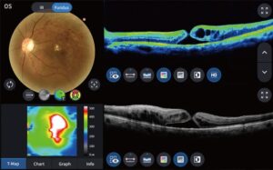
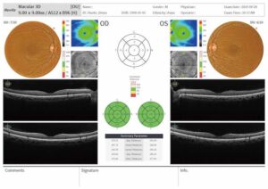
The HUVITZ HOCT-1F provides the patient’s pathology structure and relevant and essential data in an easy-to-read format and can also print the report on the analysis screen. Analysis results can be viewed via web browser and printed with different reports.
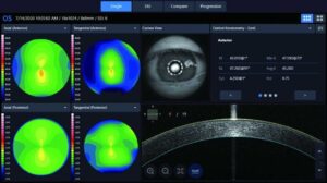
The HUVITZ HOCT-1F analyzes the anterior segment allowing measurement and analysis of corneal thickness, angle, and 3D image. Help users work more efficiently by acquiring both anterior and posterior in one place.
As a retinograph, it offers optimized color retinal images with high resolution and contrast, being very useful in clinical analysis and diagnosis. Images are provided by low flash intensity, fast capture speed, quiet operation, small pupil mode, and automatic blink detection:- High-speed, high-quality scanning.
All in one device.
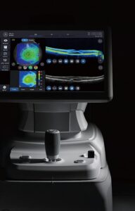
OST and fundus data are displayed. At the touch of a button, the device performs high-speed scanning and obtains high-quality images of the fundus.
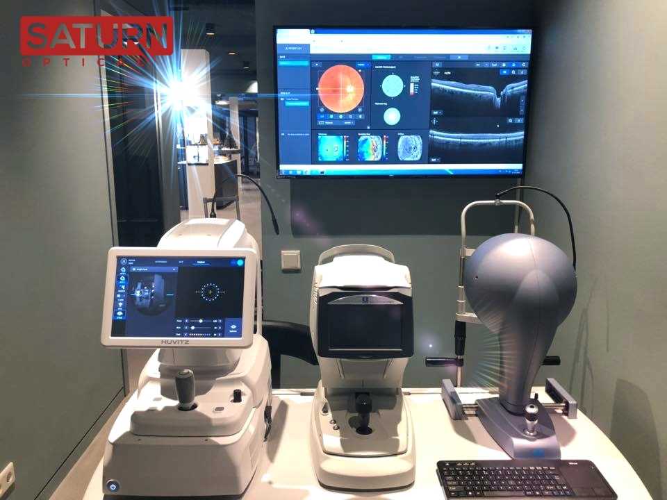
Combining a tomograph, fundus camera, angiography, and computer in one system can significantly save time and space for the user, provide user convenience, and reduce the time during the examination and diagnosis.
The patient does not need to undergo a double examination on two devices separately, which increases his comfort when visiting an ophthalmologist.
Take pictures of the fundus and perform a multimodal analysis of the retina.
A modern method of examination of blood vessels and retinal circulation – OCT-A
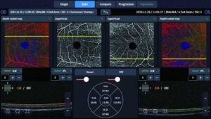
Huvitz optical coherence tomography has the function of angiography – a modern non-invasive method of visualization of vessels in the retina.
This allows you to examine the vessels in four segments of the retina: superficial, deep, external, and choriocapillaris. The size of the examination area can vary from 3×3 mm to 9×9 mm. You can also form a mosaic of different pictures. In addition, the device defines the foveolar avascular zone.
This feature is an optional option.
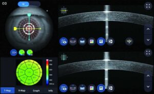
Examine the curvature of the anterior and posterior surfaces of the cornea
The software builds 16 types of maps, which provides a comprehensive examination of the patient’s cornea. This approach allows you to get the most information to choose the appropriate treatment tactics.
You can compare different data and observe the dynamics.
All necessary data on eye parameters for IOL calculation or myopia control
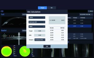
With the biometrics option, you get the basic parameters of the eye:
- FTA – the central thickness of the cornea
- ACD – depth of the front chamber
- LT is the thickness of the lens
- AL – axial length
- Also, a wide scan of the front segment makes it possible to measure:
The angle of the front camera
- Distance from white to white
- The area between the trabeculae and the iris
- The distance between the trabeculae and the iris
Examination of the cornea and anterior chamber.
The anterior segment view lens allows you to observe and measure the thickness of the cornea and the angle of the anterior chamber, thus allowing simultaneous analysis of the anterior and posterior segments.
Measuring the angle of the anterior chamber between the cornea and iris allows you to diagnose and observe the dynamics of patients with angle-closure glaucoma.
Camera viewing width – 9, 12, and 16 mm.
Huvitz HOCT-1F Specifications:
TYPE:
SC-OCT/FUNDUS EYE CAMERA
OCT:
- RESOLUTION (ON TISSUE): Z:6~7um, XY:20um
- SCANNING SPEED: 68,000A-scan/sec
- 3D ACQUISITION TIME: 1.4s(fast mode)
- MIN PUPIL SIZE: 2.5 mm in diameter
- LIGHT SOURCE: SLD 840mm
- OPTICAL POWER IN CORNEA: ≤650uW
- SCAN PATTERN (see image below table): Line (macula), cross (macula), grid (macula), radial (macula), raster (macula), 3D (macula), circle optic disc, radial optic disc, raster optical disk, 3D optical disk
FUNDUS EYE CAMERA:
- CAMERA: Color, resolution 12 mp
- FOV: 45˚
- MIN PUPIL SIZE:
- Normal: 4mm in diameter
- Small pupil: 3.3mm
- FLASH: LED
- RESOLUTION:
- Center: 60 lines/mm or more
- Medium(r/2): 40 lines/mm or more
- Medium (r): 25 lines/mm or more
COMMUN:
- WORKING DISTANCE: 33mm
- LCD SIZE: 12.1 “, resolution 1280 × 800
- DIOPTRIC COMPENSATION:
- Total Range: -33 to +33D
- -33 to -7D with a negative offset lens
- +7 to +33D without compensation lens
- SURFACE OF THE FUNDUS EYE IMAGE: NIR / Enface, FOV: 40°x30°
- INTERNAL FIXING POINT: LCD
- HORIZONTAL MOVEMENT : 70mm (forward and backward) / 100mm (left and right)
- VERTICAL MOVEMENT: 30mm
- CHIN PAD MOVEMENT: 62mm, motorized
- AUTO ALIGNMENT: X, Y for Positioning, Z for working distance
- AUTO FOCUS: Diopter adjustment for focus
- RED: DICOM File support
- DATABASE REGULATIONS: Must be configured
- EMBEDDED COMPUTER: YES
POWER SUPPLY:
AC100 ~ 240V, 50/60Hz, 1.6A~0.7A
SIZE AND WEIGHT:
- SCAN PATTERN: ACA line, radial(cornea), 3D(cornea)
- Software for analysis: layers of the cornea, thickness map, thickness and angle
ANTERIOR SEGMENT MODULE:
330(W)x542(D)x521(H)mm / 30kg
VISOR WEB:
Web access, accessible by multiple users, progression analysis, comparative analysis, 3D analysis
Include:
- Angiography software module
- Extra front segment lens 16mm
- Biometer
- Topography
What Makes Huvitz HOCT-1F Different from Other OCT Devices?
The Huvitz HOCT-1F stands out in the field of OCT devices due to its unique features, high-resolution imaging capabilities, and versatile design. Here are the key differentiators that set the Huvitz HOCT-1F apart from other OCT devices:
1. Fully Integrated Fundus Camera
The Huvitz HOCT-1F combines OCT imaging with a fully integrated fundus camera, allowing for simultaneous capture of OCT scans and fundus photographs. This integration enables comprehensive retinal assessments and enhances diagnostic accuracy by providing both structural and surface imaging in one device.
2. High-Resolution and High-Speed Scanning
The HOCT-1F offers high-speed scanning at 68,000 A-scans per second, providing high-resolution images with detailed visualization of retinal structures. This rapid scanning speed helps in reducing motion artifacts and enhances the clarity of images, making it easier to detect and monitor retinal pathologies.
3. Widefield Imaging
The device supports widefield OCT imaging, allowing for extensive views of the retina and choroid. This capability is particularly useful for detecting peripheral retinal diseases and providing a more comprehensive evaluation of the eye’s overall health.
4. Anterior Segment Imaging
In addition to retinal imaging, the Huvitz HOCT-1F includes capabilities for anterior segment imaging. This feature allows for detailed examination of the cornea, iris, and other anterior segment structures, making it a versatile tool for both posterior and anterior segment evaluations.
5. 3D Imaging and Enface Views
The HOCT-1F provides 3D imaging and enface views, which enable clinicians to visualize the retina and other ocular structures in various planes and perspectives. These imaging techniques facilitate a deeper understanding of retinal conditions and aid in the detection of subtle changes over time.
6. Automated Layer Segmentation and Analysis
The device features advanced automated layer segmentation algorithms that accurately identify and measure individual retinal layers. This capability is crucial for diagnosing and monitoring diseases such as glaucoma, diabetic retinopathy, and age-related macular degeneration with high precision.
7. User-Friendly Interface and Efficient Workflow
The Huvitz HOCT-1F is designed with an intuitive user interface and workflow enhancements that streamline the imaging process. The device includes automated functions for image capture, analysis, and reporting, reducing the time and effort required for comprehensive ocular assessments. This ease of use improves clinical efficiency and patient throughput.
8. Connectivity and Data Management
The HOCT-1F integrates seamlessly with electronic medical records (EMR) and other clinical data management systems, ensuring that patient data is easily accessible and well-organized. This integration supports efficient data management and enhances the overall quality of patient care.
9. Proven Reliability and Support
Huvitz is known for its high-quality ophthalmic equipment and strong customer support. The HOCT-1F benefits from Huvitz’s reputation for reliability and innovation, providing clinicians with a dependable tool for accurate and efficient ocular imaging.
By combining these advanced features and capabilities, the Huvitz HOCT-1F offers superior performance, versatility, and diagnostic accuracy. It is a valuable asset for eye care professionals seeking to enhance their diagnostic capabilities and provide high-quality patient care.
Reference Library
Only logged in customers who have purchased this product may leave a review.
Reviews
There are no reviews yet.