Top 10 Used OCT for Glaucoma as a Screening Tool
OCT technology was invented in 1991 to aid in the early detection and diagnosis of retinal diseases. Over the years, the Use of Optical coherence technology for glaucoma has been debated in the medical industry. Below, we explore the significant concepts of Optical Coherence Topography.
Have you been eager to learn more about the most commonly used OCT for Glaucoma as a screening tool? Then, you have come to the right place. In this article, you will learn about them and much more. However, first let’s examine the basics for a better understanding.
What is the OCT test for glaucoma?
OCT is a non-invasive imaging test that utilizes light waves to take cross-sectional pictures of the Retina. OCT reveals the Retina’s distinctive layers, enabling the mapping and measurement of their thickness. The measurements allow testing and also provide guidance for treating glaucoma.
OCT stands for Optical Coherence Tomography. OCT is a test to obtain a topographical map of the optic nerve. Non-invasive light waves used during this test take cross-sectional pictures of the retina. An OCT test will measure the nerve fiber layer. This portion of the eye is considered most vulnerable to elevated eye pressure.
Why is OCT used in glaucoma?
Optical coherence tomography for glaucoma scans will help assess all the macular regions affected by glaucoma. Assessing the macular glaucoma regions is crucial because almost 50% of the retinal ganglion cells are in the macular region. This makes macular glaucoma regions ideal for detecting any early cell loss.
What is the OCT test used for?
OCT is an imaging method that generates a picture of the back of the eye, also known as the retina. The picture measures the total dim red light reflecting off one’s retina. This makes OCT a routinely used imaging method for testing glaucoma.
How do you detect glaucoma?
To detect glaucoma, an individual requires a comprehensive eye examination. The ophthalmologist will measure the pressure of an individual’s eye to detect glaucoma. He will also inspect the drainage angle of the eye. He will also examine an individual’s optic nerve for damage and test the peripheral (side) vision. The ophthalmologist will also take a picture or the computer measurement of the optic nerve and measure the thickness of the cornea.
OCT in the Diagnosis and Management of Glaucoma

Optical coherence tomography imaging technology is used to evaluate glaucomatous structural damage. The non-invasive optical technique enables in vivo cross-sectional imaging of the ONH and retina.
The current iteration of the OCT technology, spectral-domain (SD)-OCT, has demonstrated theoretical advantages in glaucoma assessment. It offers increased axial resolution and enables faster scanning speed, leading to lower susceptibility to eye movement artifacts.
After diagnosing glaucoma through OCT, the disease can be managed by lowering the pressure on an individual’s eye. However, this situation varies from person to person, and the management options may include taking oral medications, using prescription eye drops, surgery, or laser treatment.
The Role of OCT in Glaucoma Diagnosis
OCT provides objective quantitative measurements that complement eye examination. By using OCT for glaucoma, ophthalmologists are better equipped to detect glaucoma early, as the progression of the disease can lead to irreversible loss of vision. Thus by using OCT, glaucoma can be detected before any visual loss.
Additionally, OCT also provides a baseline for identifying changes over time; furthermore, the changes on OCT predict future functional losses. It is important to note that OCT cannot be used to detect glaucoma in every patient; individuals with pre-existing conditions, among other risk factors, as we will elaborate on later, do not require OCT to diagnose glaucoma. However, OCT might still be needed to detect progression.
Images from the OCT scans are usually examined from top to bottom and left to right within eight steps. The interpretation of SD-OCT images encompasses various aspects to ensure accurate detection of the condition.
How Does Glaucoma Happen
When fluid builds up in the front part of the eye, it causes an increase in eye pressure, subsequently damaging the optic nerve. And you will not want your optic nerve damaged because it carries information from your eye to the brain. Thus once the optic nerve is damaged, it could lead to serious eye problems.
Importance of Carrying Out Glaucoma Testing
A couple of tests are typically performed to diagnose glaucoma. The advantage of conducting the tests early is that if glaucoma is diagnosed promptly, the individual can take steps to prevent vision loss.
How Many Types Of Glaucoma Are There?
There are several types of Glaucoma; we will, however, look at the main types of glaucoma, the first one being
Closed-angle glaucoma
closed-angle glaucoma has two aka’s and can be referred to as narrow-angle glaucoma or angle-closure glaucoma. It is a type of glaucoma that affects one eye at a time; if an individual gets this type of glaucoma, their drainage canals will be covered. The above is why closed-angle glaucoma can be categorized as acute or chronic.
Individuals who suffer from acute closed-angle glaucoma will be subjected to a rapid increase in eye pressure. The individuals could lose their vision in hours if the condition is not treated promptly. Acute angle glaucoma is therefore categorized as a medical emergency.
Chronic closed-angle glaucoma is known to develop slowly; the individual will not experience any symptoms up until the damage is severe.
The second primary type of glaucoma is open-angle glaucoma, also called primary open-angle glaucoma.
Many people have been found to suffer from this type of glaucoma, which usually occurs when the eyes cannot drain fluid properly via the eye’s drainage canals. The fluid then gets backed up in the canals, consequently causing an increase in eye pressure.
Open-angle glaucoma has been established to develop slowly and can ultimately take years or months to manifest. Individuals who suffer from this glaucoma never have any symptoms or vision changes at the beginning. But once infected, open-angle glaucoma will present in both eyes simultaneously.
Another type of glaucoma that people must be familiar with is normal-tension glaucoma, which does not fall under the two main types. Thus, people with this type of glaucoma tend to experience eye pressure within the normal range but still show signs of glaucoma. Individuals suffering from normal-tension glaucoma might experience optic nerve damage or have blind spots in their field of vision.
Importance of Glaucoma Testing
As mentioned earlier, individuals who suffer from open-angle glaucoma may not exhibit any symptoms until the disease has developed into a more serious stage. And that is why individuals who have certain risk factors must be tested. The above brings us to the next big question what are some of the risk factors of glaucoma?
Individuals with glaucoma in their family history are at risk of developing complications. Glaucoma has also been found to affect older people; thus, individuals aged 60 and above are at risk of developing eye complications. Also, Hispanics aged 60 and above have a higher risk of developing glaucoma when compared to native Europeans.
African Americans are also at a greater risk of developing glaucoma, while individuals of Asian descent have a much higher risk of developing closed-angle glaucoma. Individuals who have had eye injuries are at risk of developing glaucoma. The same applies to people who are far-sighted and near-sighted.
The long-term use of steroid medication can lead to the development of glaucoma. Individuals with conditions such as high blood pressure, diabetes, and migraines, among other health problems that affect the entire body, are at a high risk of contracting glaucoma.
What Is a Comprehensive Eye Exam in Glaucoma Testing?

Given the severity of the two main types of glaucoma, it is natural to want to know the eye exam that an ophthalmologist is likely to carry out. Below is an outline of the exams carried out by an ophthalmologist when testing for glaucoma.
Tonometry
the tonometry test will require an individual to sit in an exam chair next to a slit lamp (special microscope). The ophthalmologist will then numb the eyes using eye drops, and the individual will be required to rest their chin and forehead on the slit lamp. And while leaning into the slit lamp, the eye specialist will use a tonometer to measure eye pressure. During this examination, the individual will experience a small puff of air but nothing painful.
Pachymetry
– when performing the tonometry test, the first step involves numbing the eyes using eye drops. The eye specialist will then use the pachymetry device to measure the cornea’s thickness, which is known to predispose individuals to a higher risk of developing glaucoma.
Perimetry
is better referred to as the visual field test and is used to measure peripheral (side) vision. Under the perimetry test, an individual must stare straight ahead at a screen. A light or image will then move in from one side of the screen. The patient’s job is to let the eye specialist know when they detect the light or image while looking straight ahead.
Dilated eye test
when conducting this test, the specialist will first put eye drops into the eyes, dilating the pupils. With a device with a light and a magnifying lens, he/she will examine the optic nerve to check for damage.
Gonioscopy
under this test, the ophthalmologist will put drops in the patient’s eyes to numb and dilate them. He/she will then place a special handheld contact lens on the patient’s eyes. The lens is typically designed with a mirror, allowing the specialist to view the inside of the eye from various angles. The lens can show if the angle between the iris and the cornea is too wide, indicating the possibility of open-angle glaucoma. And if the angle is too narrow, it would be a possible sign of closed-angle glaucoma.
How to Prepare For a Glaucoma Test
If you have never done any eye test, it is not uncommon to wonder what might transpire. What you need to know is that when your eyes are dilated, your vision may become blurred, and you may become more sensitive to both natural and artificial light. This particular condition might persist for a few hours, and the severity will vary from person to person.
To be safe and protect your eyes from bright light, consider investing in sunglasses, which you can wear after your appointment with the ophthalmologist. Most importantly, you may not be able to drive yourself back home, and as such, you should bring along a company that will drive you back home, because your vision will be too impaired to enable you to drive safely.
Are there any risks associated with the Glaucoma Test?
There haven’t been any known risks associated with Glaucoma testing; however, some of the tests described above could be uncomfortable and temporarily blur your vision.
Can the Damage Caused By Glaucoma Be Reversed?
No, the damage caused by glaucoma cannot be reversed, which means the condition will be permanent. However, further damage can be halted through surgery and the prescribed medicine.
Can OCT Diagnose Other Conditions?
Apart from glaucoma, OCT can also diagnose diabetic retinopathy, vitreous traction, macular edema, macular pucker, macula hole, central serous retinopathy, vitreous traction, and age-related macular degeneration. OCT has been instrumental in diagnosing optic nerve disorders, but it cannot be relied upon to diagnose conditions that interfere with light passing through the eye.
What Follows If Diagnosed With Glaucoma?
Given the outcome of the tests and the determination that you indeed suffer from glaucoma, one or more of the following treatments will be recommended.
The Ophthalmologist may recommend that you undergo surgery, creating a new opening that will enable the fluid to leave the eye. The specialist might also recommend certain medications, which can lower an individual’s eye pressure or even cause the eye to produce less fluid. Regarding prescriptions, some medicines may be in the form of pills, while others are in the form of eye drops.
The ophthalmologist could also recommend the implantation of a drainage tube, a type of surgery where a flexible plastic tube will be placed in the eye to help drain the excess fluid.
Here are the Top 10 commonly used OCTs for Glaucoma as a Screening Tool.
1. Optovue Avanti ($19,999.00)

Optovue Avanti is a wide-field device OCT. This test provides a multi-layered assessment of peripheral pathology at various levels of the choroid and retina. Thus, a new standard for OCT imaging was established.
A few Features of Optovue Avanti:
- Wide Field En-Face OCT: Eyecare practitioners can use Optovue Avanti OCD to avoid all clinical challenges as it uses the most current technology.
- Establishes New OCT Imaging Standards: Optovue Avanti offers En Face OCT with a 40° wide field. The OCT test will assess peripheral pathologies giving rise to advanced OCT platforms.
- Motion Correction Technology data proprietary algorithms applied by the motion correction technology help reduce artifacts caused by motion eye motion while scanning.
- 2-Phase Noise Reduction: The 2-Phase noise reduction feature helps reduce signal noise during scan acquisition and during post-processing data capture.
- Deep Choroidal Imaging: The deep choroidal imaging feature or DCI helps push signal strength into the retina’s choroidal area with a button.
- Retina Module: The feature will enable specialists to assess the retina’s parts and evaluate change over time.
2. Optovue iFusion ($8,888.00)

Fundus and OCT imaging are made possible in one instrument with the Optovue iFusion. This tool combines the best of Fundus imaging and Spectral-Domain OCT.
Features of Optovue iFusion:
- Complete Solution For Retinal Imaging: Optovue iFusion is the complete solution for retinal imaging as its modularity provides numerous upgrade pathways from which one can choose.
- Range Of Retinal Scans: This tool helps take a range of OCT retinal scans such as 3D retina cube, real-time en face, retina map, and radial line.
- Extensive Scans For Glaucoma: Some extensive OCT and more offered by iFusion include the following:
- 3D Disc Cube: In-depth analysis of one’s optic disc structure is possible with iFusion’s 3D disc cube scans.
- Nerve Fiber Layer Analysis: Quantification and visualization of RNFL thinning in glaucoma patients can be performed using a nerve fiber thickness map.
- Ganglion Cell Complex Analysis: Ganglion cell loss due to glaucoma can be measured and identified using GCC thickness map analysis, which is delivered by this tool.
- Comprehensive Reports: A Comprehensive analysis of the patient can be done using the combo, GCC, and RNFL analysis reports.
3. Wavelight Topolyzer Vario ($19,999.00)

Wavelight Topolyzer Vario diagnostic device helps take precision measurements, enabling doctors to offer personalized treatments. From navigating corneal scans to viewing and measuring them, one can do it efficiently with just a few clicks.
Features of Wavelight Topolyzer Vario:
- Keeps It Simple: This diagnostic device has a refined navigation system, a one-click printing feature, intuitive image selection pupil detection, simplified drop-down menus, smooth export functions, and WaveNet™ integration.
- Easily Compare Scans: One can easily compare scans through automatically ordered exams, side-by-side on-screen positioning, and ease of switching between ‘excluded’ and ‘preferred’ scans.
- Helps Perform Advanced Diagnostics: Advanced diagnostics is possible with the advanced Vario platform, Contoura® Vision. Thus providing a more personalized and satisfactory experience to all the engaging patients.
- Get the Whole Picture: This navigation device enables the visualization of the entire picture of a glaucoma patient’s eye. This device’s scan covers the screen more than the previous version. It provides one-click toggles between topography, camera, and Placido imaging views.
4. Nidek Mirante ($25,999.00)
Nidek Mirante is the ultimate multimodal imaging platform with a state-of-the-art OCT or SLO combo. The tool offers ultra-wide HD images and FlexTrack technology.
Features of Nidek Mirante:
- Unsurpassed Clarity & Color: Nidek Mirante offers unsurpassed clarity and color for every detail. One can make detailed pathology evaluations using its 163 ° ultra-wide-field color image in refine mode. The tool also provides panorama image composition.
- FlexTrack Technology: The new FlexTrack technology can enhance average image quality. This helps doctors correct a distorted image caused by poor fixation and correct it using this technology.
- Simple Interface & Easy Operations: The Nidek Mirante tool has multiple functions and modalities. However, the unique interface software of this testing tool makes it pretty simple and easy to use. Presenting all multimodal images directly on the summary screen enables more comprehensive and faster disease evaluation.
- Glaucoma Analysis: This Mirante tool also incorporates maps with thicknesses ranging from 16.5 to 12mm. This map clearly and visually represents all the significant pathological changes from the periphery to the central retina.
- Tilt & Swing Features: The optical head of this tool features tilt and swing capabilities, enabling it to capture images of the fundus periphery. This feature also helps the tool acquire panorama patients. Thus, allowing all those patients with unstable fixation pretty much.
5. Optopol REVO FC ($12,999.00)

Optopol REVO FC is a real-time eye-tracking hardware device. It is an all-in-one fundus and OCT camera. This tool features a comprehensive suite of capabilities, including accurate fundus color imaging, high-resolution hardware tracking OCT, and more.
Features of Optopol Revo FC
- Fundus Camera With OCT Functionality: This all-in-one device with OCT technology is one compact system that offers high-quality and detailed OCT images with detailed fundus photos, enabling multipurpose diagnosis.
- Offers Proven REVO System Advantages: This device provides proven REVO system advantages with cutting-edge color fundus imaging. Diagnostic certainty becomes achievable with high-quality, comprehensive OCT scanning analysis of the eye’s retinal layer.
- All-In-One Device: This OCT technology-rich device is a complete, all-in-one system featuring a full-color fundus camera within a single, compact unit. Its ability to capture fundus color images and OCT scans in a shot makes it a significant space saver.
- New Linking Function: The new linking feature in this device enables a single fundus photo to be linked to multiple OCT exams. This helps to reduce the total shots number per eye significantly.
6. ZEISS CIRRUS 6000 ($49,999.00)
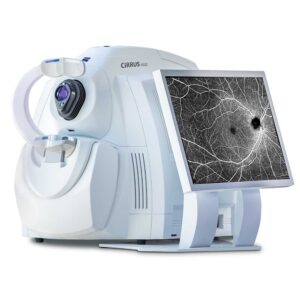
Zeiss Cirrus 6000 is another OCT for glaucoma testing tool that can deliver up to 100,000 scans in one second. This next-generation OCT device from ZEISS can deliver high-speed image capturing and HD detailed imaging. Thus helping one make more informed decisions.
Features of ZEISS CIRRUS 6000:
- Faster & Wider Level of Detailing: ZEISS CIRRUS 6000 enables clinicians to capture over 100,000 scans in under one second. It captures high-definition OCT scans and OCT Angiography scans. Thus, providing them with more insights into the patient’s condition.
- Makes Revolution Routinely: The ZEISS AngioPlex OCT Angiography helps facilitate non-invasive imaging of the retina’s microvasculature. This aids in taking retinal and glaucoma disease management and treatment to the next level by assessing and analyzing a good range of pathologies.
- CIRRUS-Powered Treatment Decisions: This CIRRUS-powered OCT technology device provides clinically validated and extensive applications for glaucoma, anterior, and retina segments. Thus, it enables faster throughput, precise analysis, and more informed decision-making.
7. ZEISS PLEX Elite 9000 ($30,999.00)

ZEISS PLEX Elite 9000 is a swept-source OCT Angiography device that aims to cover the undiscovered. This device uses transformational imaging OCT technology. Thus helping glaucoma and retina researchers get microvascular and structural clarity.
Features of ZEISS PLEX Elite 9000:
- Swept Source Dual-Speed OCT: Deeper, faster, higher resolution, and dual-speed new PLEX Elite 9000 takes clinical research and ophthalmic imaging to the next level. Clinicians can utilize the information obtained from this dual-speed OCT device to investigate any clinical benefits acquired at a scan speed of 200kHz.
- Helps Explore Deeper Meanings: Exploring deeper meanings is made possible with the ZEISS PLEX Elite 9000. This device helps clinical researchers see the potentialof the disease profoundly and widely. This device examines retinal progression to assess the response of the choroid and retina. This device can even explore choroidal pathology.
8. Optos Silverstone ($20,555.00)

Optos Silverstone is one of the most powerful tools for examining one’s retina. This ultra-widefield imaging retinal has been integrated with swept-source UWF-guided optomap images.
Features of Optos Silverstone:
- Combines UWF & SS-OCT: The Optos Silverstone device combines the optimal-guided OCT swept source with ultra-widefield or UWF fundus capture. Thus aiding in the improved management of patients and making impactful treatment decisions.
- Significant Peripheral OCT Imaging: This device can capture peripheral pathologies using Optomap integrated imaging and swept-source full-field imaging. This usually enables clinicians to get anatomical and guided media insight essential for crucial surgery management. Thus, providing significant clinical peripheral OCT imaging.
- Direct Contribution to Plans for Patient Management: In a few cases, the navigated Optomap SSC-OCT aids significantly when it comes to patient management plans, such as surgical, laser, or injection treatments.
9. Ziemer Galilei G6 ($15,555.00)

Galilei G6 combines the benefits of Optical Biometry, Tomography, and Topography. The device provides access to the total corneal wavefront, pachymetry, astigmatism data, and curvature. Thus, providing surgeons with a complete data set for planning cataract and refractive surgery.
Features of Galilei G6:
- All-In-One Unit: This device is an all-in-one unit that combines the benefits of Scheimpflug tomography, optical biometry, and Placido–disc–based topography.
- Offers Comprehensive Screening: The aforementioned all-in-one combination enables Galilei G6 to provide comprehensive screening for refractive surgery or cataracts, providing complete data.
- Improves Work Maintenance, Efficiency, & Optimisation: This device helps store and gather data on a single device. Thus, improving the clinician or practitioner’s workflow efficiency, reducing maintenance costs, and optimizing office space utilization.
- Complete Data Set For Planning & Surgery: This device provides surgeons access to the total corneal wavefront, high-definition pachymetry, astigmatism data, and curvature. Thus helping medical practitioners plan for cataract surgery.
10. Canon Xephilio oct-a1 ($15,999.00)

One device taking OCTA to the next level is the Canon Xephilio OCT-A1 device. This device enables the rapid and easy acquisition of essential data, providing incredible detail.
Features of Canon Xephilio oct-a1
- New Retinal Imaging Reality: The Canon Xephilio OCT-A1 device offers multiple operational options, enabling safe patient examination.
- Outstanding Imaging Quality: Due to Canon’s recognized expertise in optics, one can expect superb image quality from this device. Thus enabling excellent individual layers and structure differentiations of the retina.
- Consistent & Automatic High-Quality Images: In most cases, involuntary eye movement becomes unavoidable during examinations. However, the device’s integrated real-time SLO retinal tracking maintains the same position during scanning and provides automatic results.
Conclusion
Optical coherence tomography (OCT) is a valuable clinical tool for diagnosing and detecting glaucoma progression in patients. Getting an OCT for glaucoma done using the correct method will provide accurate information for diagnosing glaucoma. From detecting moderate glaucoma to late disease conditions, early detection of glaucoma is certainly possible with the best OCT test tool.
Of the two types of glaucoma, closed-angle glaucoma requires immediate treatment because it can potentially cause blindness. Some symptoms that individuals should, therefore, watch out for include severe eye pain, red eyes, sudden blurring of vision, colored halos around lights, nausea, and vomiting.
Once diagnosed with glaucoma, the ophthalmologist will want to monitor your vision regularly and ensure that the recommended treatment prevents additional vision loss. It is essential to note that glaucoma cannot be cured, and the treatment measures instituted will not restore vision. However, if detected early, significant vision loss can be prevented.