Topcon 3D OCT 1- Maestro 1 – OCT and Fundus Camera with Retinal Camera
Original price was: $14,999.00.$7,999.00Current price is: $7,999.00.-47% OFF
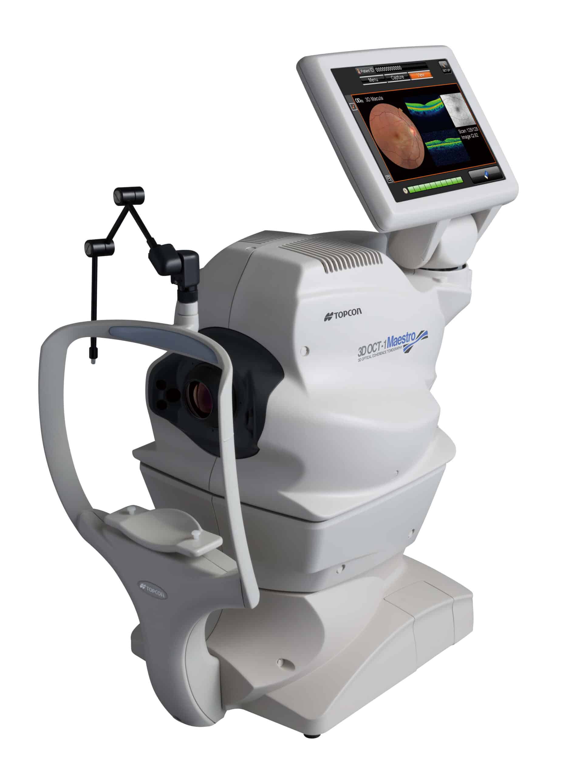
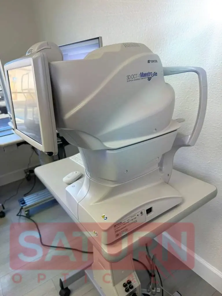
The Topcon 3D OCT-1 Maestro (Maestro 1) is a combined OCT and fundus/retinal camera designed to speed up everyday imaging in optometry and ophthalmology settings. Capturing cross-sectional OCT scans alongside color fundus images in a single workflow supports efficient evaluation and follow-up for glaucoma, macular conditions, and routine retinal documentation.
In stock
Description
Topcon 3D OCT 1- Maestro 1 sets a new standard for clinical usability by combining a high-resolution, mydriatic-free color retinal camera with the latest Spectral Domain OCT technology.
The Topcon 3D OCT-1 Maestro Retinal Camera is a high-performance medical device, specially designed for
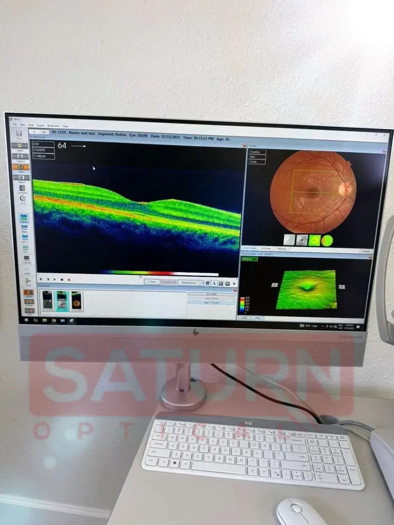
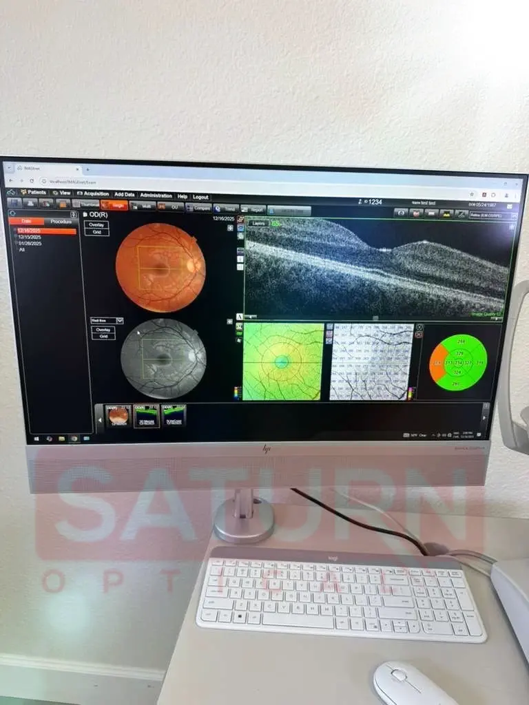
retinal imaging by providing technology. The latter combines an optical coherence tomography (OCT) scanner and a color camera to provide 3D images of the retina, enabling a thorough analysis of ocular health.
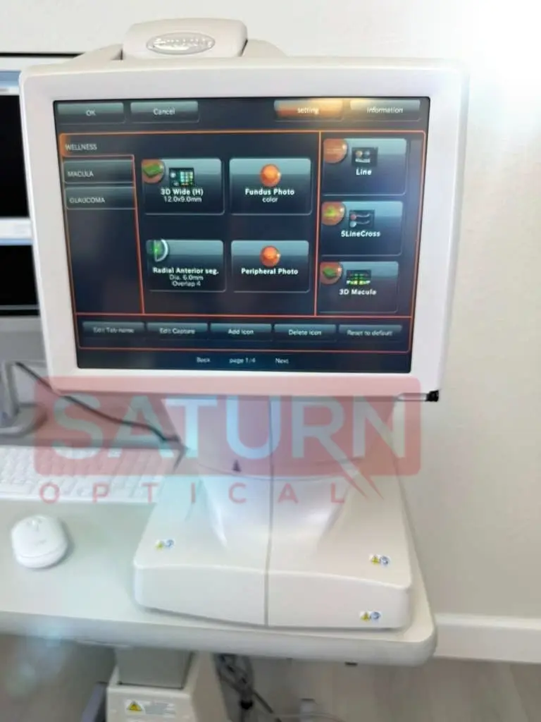
The 3D OCT-1 Maestro also offers multiple imaging modes, including single-point and 3D scanning, for comprehensive analysis of the retina. In addition, its wireless connectivity makes it easy to share data and images with other healthcare professionals.
Topcon 3D OCT 1- Maestro 1 Features
- Full automation (alignment, focus, and capture) 3D OCT and fundus capture technology, combined with automatic report functions, delivers a fast office workflow and ensures that any of your staff can easily take images
- Comprehensive assessment of ONH and macula in one scan with an innovative 12mm x 9mm scanning protocol
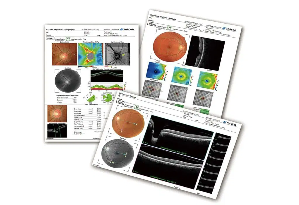
- Extensive Reference Database of RNFL, Total Retinal, GCL + IPL, and GCL + IPL + RNFL thickness measurements
- 50,000 A Scans/sec Spectral Domain OCT with a non-mydriatic color fundus camera saves space and expense of purchasing 2 systems
- High quality images – 2D, 3D and fundus images
- PinPoint™ Registration of the OCT image with the fundus image
- Latest automatic layer segmentation algorithms
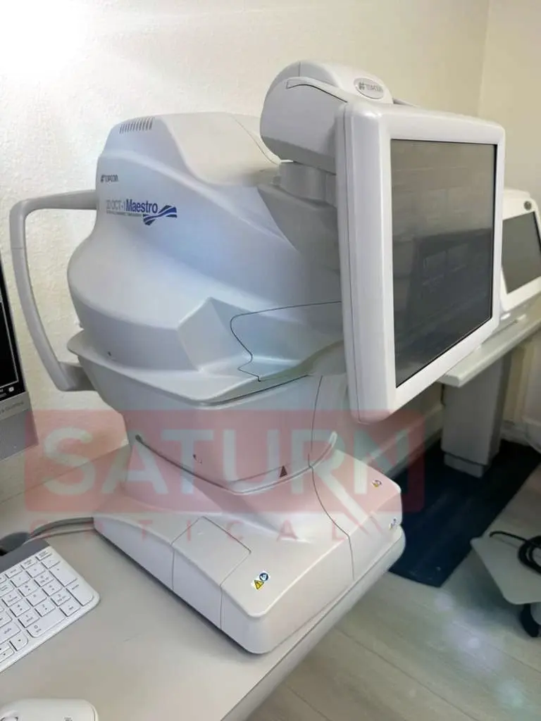
- All-in-one compact system to fit small office settings
- An online instrument training program significantly reduces the training time of new staff
The system boasts rich analysis and reporting functions, empowering clinicians with comprehensive insights into ocular health. From EN VIEW OCT Imaging to true color fundus photography, the Maestro 3D OCT-1 delivers unparalleled imaging capabilities, enabling detailed examination of the retina and surrounding structures with superb OCT technology.
In addition to its advanced imaging capabilities, the Maestro 3D OCT-1 features a compact and space-saving design, making it ideal for any clinical setting. Its network and DICOM connectivity further enhance its versatility, allowing for seamless integration into existing practice workflows.
The system offers both full-auto and semi-auto capturing modes, providing flexibility to suit varying clinical scenarios. With semi-auto capturing, operators can easily initiate scans at their convenience, while the live fundus view feature facilitates imaging of small pupils with clarity and precision.
Furthermore, the Maestro 3D OCT-1 includes specialized modes, such as cataract mode, which adjusts scan positions to accommodate cataract-induced opacity, ensuring clear and accurate imaging even in challenging cases. Topcon’s unique stereo-matching automatic alignment technology further enhances efficiency and stability, enabling quick and reliable alignment for every scan.
Advantages:
- Fully automatic OCT with a simple touch of a finger,
- Rich analysis and reporting features,
- Reliable scanning assistance,
- High-quality, high-resolution OCT image and color fundus image,
- Seamless network solution,
- Compact size and flexible layout,
- Fully automatic capture,
WHAT’S INCLUDED
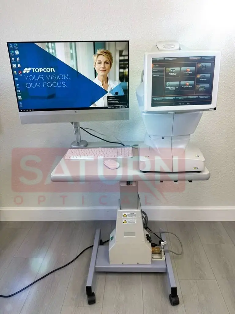
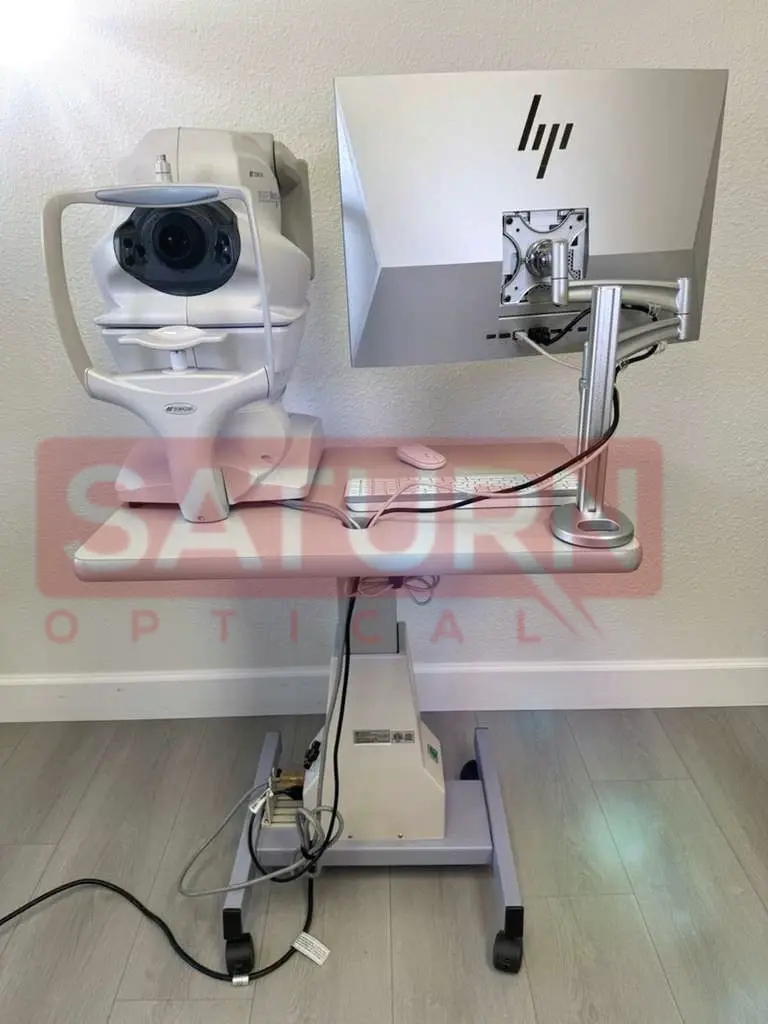
- All-in-One Computer
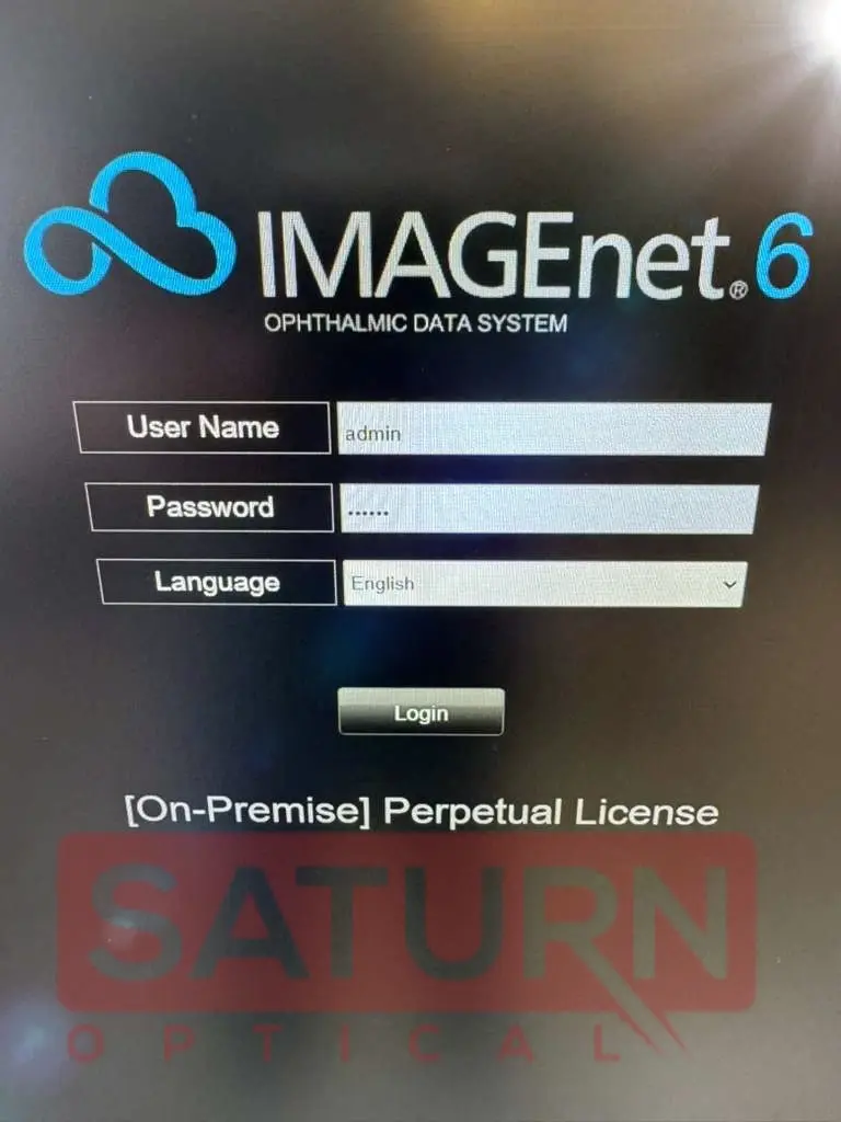
- Windows 10 + Imagenet R6 Software
- Table
- Manual (USB Drive)
- All required accessories
- Professional packaging
- Fully tested and calibrated
- Ready for clinical use
- Integrated retinal camera
SPECIFICATIONS
- Observation & Photography of Fundus Image
- Scan Mode: Color, Red-free (display digital red-free)
- Picture Angle: 45°/30° or equivalent (digital zoom)
- Operating Distance
- 8 mm (in fundus photography)
- 6 mm (in anterior segment photography)
- Photographable Diameter of Pupil
- 45°: Ø4.0 mm or more
- Small pupil diameter: Ø3.3 mm or more
- Scan Range
- On fundus
- Horizontal direction 3 – 12 mm
- Vertical direction 3 – 9 mm
- On cornea
- Horizontal direction 3 – 6 mm
- Vertical direction 3 – 6 mm
- Scan Speed: 50,000 A-Scans per second
- Lateral Resolution: 20 µm
- In-depth Resolution:6 µm
- Photographable Diameter of Pupil: Ø2.5 mm or more
- Internal Fixation Target: Dot matrix type organic EL (The display position can be changed and adjusted. The presenting method can be changed.)
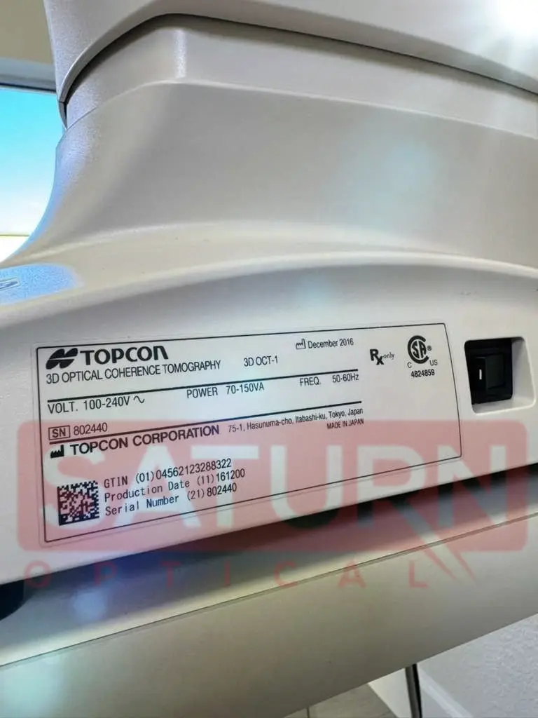
- Electric Rating
- Source Voltage: AC 100 – 240V
- Power Input: 70 – 150VA
- Frequency: 50HZ – 60HZ
- Dimensions & Weight
- Dimensions: 1” – 17.4” (W) x 18.5” – 26.3” (D) x 20.4” – 28.4” (H)
- Weight: 3 lbs.
Reference
Only logged in customers who have purchased this product may leave a review.
Reviews
There are no reviews yet.